AI Radiology Image Analysis Tools: Uses, Benefits, Risks and How to Choose
A practical 2026 guide for radiology leaders, clinicians, healthcare operators and technology buyers
| Editorial note: This article explains how to evaluate AI-assisted imaging software. It does not provide medical advice, endorse a particular vendor or replace clinical judgment, local validation, regulatory review or professional oversight. |
AI Radiology Image Analysis Tools: What They Do and How to Choose
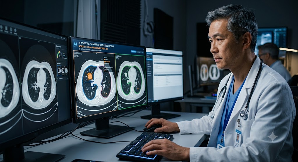
Radiology generates a constant stream of complex images—from chest X-rays and mammograms to CT and MRI studies. AI radiology image analysis tools are designed to help clinicians manage that volume by analyzing pixels, identifying patterns and returning structured outputs inside or alongside the reading workflow.
The important word is help. Most clinical imaging AI is assistive: it may flag a suspected abnormality, quantify a lesion, prioritize an urgent study or act as a second reader. The radiologist remains responsible for interpreting the full examination in its clinical context, subject to the product’s intended use and local rules.
This guide explains the main tool categories, representative platforms, realistic benefits, common limitations and the questions healthcare organizations should ask before buying or deploying a solution.
What are AI radiology image analysis tools?
AI radiology image analysis tools are software systems that apply machine-learning methods—often deep learning—to medical images or related imaging data. Depending on the product, they can detect, classify, segment, measure, compare or prioritize findings. Some products address one narrow task; others combine multiple algorithms or route third-party models through a shared platform.
Typical inputs include DICOM images from X-ray, CT, MRI, ultrasound or mammography. Outputs may appear as overlays, heatmaps, measurements, probability scores, structured results, worklist changes or notifications. A useful implementation connects those outputs to the radiologist’s existing PACS, RIS, reporting and communication workflow instead of creating another isolated screen.
Common categories of radiology AI software
Detection and computer-aided diagnosis: Flags patterns associated with conditions such as intracranial hemorrhage, pulmonary embolism, pneumothorax, fractures, lung nodules or breast lesions, within the product’s cleared or approved intended use.
Triage and worklist prioritization: Moves studies with suspected time-sensitive findings higher in the reading queue or alerts designated care-team members. Triage is not the same as a final diagnosis.
Segmentation and quantification: Outlines anatomy or lesions and calculates measurements such as volume, density, burden or change over time.
Screening support and second reading: Provides an additional assessment in high-volume screening programs such as mammography or chest imaging.
Image quality and acquisition support: Helps with positioning, reconstruction, protocol consistency, dose optimization or image-quality checks.
Workflow orchestration: Routes studies to appropriate algorithms, consolidates results and helps IT teams manage integrations, versions and operational monitoring.
Representative AI radiology image analysis tools
The following examples illustrate different product approaches. Availability, indications, regulatory status and product names vary by country and can change. Verify every claim against the current regulator database and the vendor’s official labeling before clinical use.
| Example | Primary approach | What it is designed to support | Potential fit |
| Aidoc | Enterprise clinical AI platform | Imaging analysis, worklist prioritization, care-team activation and follow-up workflows across multiple use cases. | Organizations seeking a platform approach rather than a single point solution. |
| Harrison.ai / Annalise | Comprehensive decision support | Chest X-ray and CT-focused tools designed to identify multiple findings and support prioritization or review. | Sites comparing broad multi-finding coverage within specific modalities. |
| Gleamer | Musculoskeletal and X-ray assistance | Products centered on fracture detection and related radiography workflows, with additional imaging applications in its portfolio. | High-volume emergency, trauma or general X-ray environments. |
| Lunit INSIGHT | Chest and breast imaging AI | CXR, mammography and tomosynthesis support, including lesion highlighting and screening assistance. | Programs focused on thoracic or breast imaging workflows. |
| Qure.ai | Imaging detection and workflow support | Chest X-ray and head CT applications, including analysis and prioritization use cases. | Health systems evaluating targeted imaging AI across diverse care settings. |
| Viz.ai / RapidAI | Time-sensitive care coordination | Imaging-based detection or triage combined with notifications and clinical workflow coordination, especially in acute-care pathways. | Stroke and other time-critical service lines where communication speed is central. |
Note: This is a representative, non-ranked list—not a complete market directory. Product capabilities and legal availability must be checked for the specific geography, modality, patient population and intended workflow.
How these tools fit into a radiology workflow
Image acquisition: The scanner or modality sends the study to the imaging environment, usually in DICOM format.
Routing: Rules determine whether the study is sent to one or more approved AI algorithms.
Inference: The algorithm analyzes eligible images and returns its output, ideally with status and error handling.
Presentation: Results appear in the PACS viewer, worklist, reporting application or a tightly integrated companion interface.
Clinical interpretation: The radiologist reviews the complete study and clinical context, considers the AI output and records the final interpretation.
Monitoring: The organization tracks uptime, latency, concordance, subgroup performance, overrides, user behavior and changes in workflow or model version.
| Key takeaway: A high-performing algorithm can still fail operationally if it arrives late, interrupts reading, generates too many false alerts or does not integrate with existing systems. Workflow fit is part of clinical performance. |
Potential benefits
Faster prioritization of studies that may contain time-sensitive findings.
More consistent measurements and quantitative follow-up across serial exams.
A second set of eyes for selected, well-defined tasks.
Reduced manual work when outputs populate structured fields or trigger approved workflows.
Better visibility into incidental or follow-up findings when the product and workflow are designed for that purpose.
Operational data that can help teams identify bottlenecks and quality-improvement opportunities.
These are potential outcomes, not guarantees. The effect depends on baseline performance, prevalence, case mix, local population, integration, thresholds, alert design and how clinicians actually use the output.
Risks and limitations to plan for
False positives and false negatives: AI can flag a finding that is not present or miss one that is. Sensitivity and specificity must be interpreted alongside prevalence and clinical consequences.
Dataset shift: Performance may change when scanners, protocols, demographics, disease prevalence or referral patterns differ from the development data.
Automation bias: Users may over-trust an AI suggestion or spend disproportionate effort resolving disagreements with it.
Alert fatigue: Too many low-value notifications can slow teams and make genuinely urgent alerts easier to overlook.
Integration and downtime: Broken routing, delayed results, incomplete DICOM handling or interface failures can create new safety risks.
Equity and subgroup performance: Aggregate accuracy can hide weaker performance in particular demographic or clinical groups.
Privacy and cybersecurity: Medical images and metadata are sensitive health data; cloud architecture, access controls, retention, audit logs and incident response matter.
Regulatory mismatch: A product authorized for one intended use, modality or population should not be treated as approved for every adjacent task.
How to choose the right AI radiology tool
Start with a clinical and operational problem—not a product demo. A focused use case makes it possible to define success, compare alternatives and stop a pilot that does not add value.
1. Define the use case
Specify modality, body region, patient population, care setting, workflow stage and intended user. Decide whether the goal is detection, triage, quantification, screening support or orchestration.
2. Verify regulatory status
Check the current regulator database and official labeling for the exact product version, indication and geography. In the United States, use the FDA’s AI-enabled medical-device list and linked decision records as a starting point.
3. Examine clinical evidence
Look for external validation, multi-site data, representative case mix, appropriate reference standards and performance reported with confidence intervals—not only a headline accuracy number.
4. Test locally before go-live
Run acceptance testing on representative local cases and workflows. Evaluate failure modes, subgroups, scanners, protocols and edge cases relevant to your site.
5. Assess integration
Confirm DICOM, HL7 and FHIR requirements; PACS/RIS/reporting compatibility; routing logic; latency; result display; single sign-on; audit trails; downtime behavior; and support ownership.
6. Evaluate human factors
Observe radiologists and technologists using the tool. Measure whether it reduces or adds clicks, changes attention, affects reading time or creates alert fatigue.
7. Build the economic case
Include licensing, infrastructure, interfaces, cybersecurity review, validation, training, monitoring, support and model-change management—not just subscription price.
8. Plan continuous monitoring
Set baselines, owners, review cadence and thresholds for investigation. Track performance drift, discordance, latency, failures, subgroup variation and user overrides.
A practical evaluation scorecard
| Dimension | Question to answer | Score (1–5) |
| Clinical value | Does it address a high-priority problem with measurable patient or workflow impact? | ____ |
| Evidence | Was it validated externally and on populations similar to ours? | ____ |
| Safety | Are failure modes, contraindications, uncertainty and escalation paths clear? | ____ |
| Workflow fit | Does the result appear at the right moment in the tools clinicians already use? | ____ |
| Interoperability | Can it integrate reliably with current and planned systems using maintainable standards? | ____ |
| Governance | Can we inventory, version, audit, monitor and retire it responsibly? | ____ |
| Security and privacy | Do architecture, data flows, access controls and contracts meet organizational requirements? | ____ |
| Total cost | Is the expected benefit credible after implementation and ongoing operational costs? | ____ |
Implementation roadmap
Form a multidisciplinary governance group with clinical, technical, security, legal, quality and operational representation.
Create a documented baseline for the target workflow before introducing AI.
Shortlist products using intended use, evidence, integration and support criteria.
Complete security, privacy, regulatory and contracting reviews.
Conduct local acceptance testing and a controlled pilot with predefined success and stop criteria.
Train users on intended use, limitations, failure modes, downtime and reporting of concerns.
Go live gradually, monitor closely and retain a clear rollback path.
Review performance regularly and reassess after software, scanner, protocol or population changes.
Frequently asked questions
Can AI read radiology images without a radiologist?
Some products may perform narrowly defined autonomous functions where permitted, but most radiology AI tools are designed to assist qualified clinicians. The product’s intended use, regulatory status and local policy determine how it may be used.
Which imaging modalities can radiology AI analyze?
Products exist for X-ray, CT, MRI, mammography, ultrasound and other imaging data. Coverage varies widely by tool, anatomy, acquisition protocol and clinical indication.
Are AI radiology tools accurate?
Many tools show strong performance for specific tasks, but accuracy is not one universal number. Evaluate sensitivity, specificity, predictive values, calibration, confidence intervals, external validation and local performance for the relevant population.
Will AI replace radiologists?
The more immediate pattern is task-level augmentation: prioritization, detection support, segmentation, measurement and workflow automation. Radiologists integrate imaging findings with history, prior studies, uncertainty and clinical consequences—responsibilities that extend beyond a single algorithm output.
How do hospitals verify an AI imaging tool?
They should confirm regulatory status and intended use, review evidence, perform local acceptance testing, validate integration and human factors, document governance and monitor real-world performance after deployment.
What is the biggest mistake when buying radiology AI?
Buying a technically impressive model without a defined workflow problem, measurable success criteria, local validation or a plan for ongoing monitoring.
SUGGESTED EXCERPT
AI radiology tools can flag findings, prioritize worklists and support quantitative analysis—but results depend on fit, integration and continuous monitoring. Here is how to evaluate them responsibly.
Final thoughts
AI radiology image analysis tools can support detection, triage, quantification and workflow coordination. Their real value, however, is determined after the demo: in the fit between the tool, the patient population, the radiologist, the imaging systems and the organization’s safety processes.
The strongest buyers treat imaging AI as a clinical system with a lifecycle—not a one-time software purchase. They define a narrow problem, verify evidence and regulatory status, test locally, integrate carefully and monitor continuously. That approach creates a better chance of improving care without introducing invisible operational risk.
Sources and fact-check links
U.S. FDA — Artificial Intelligence-Enabled Medical Devices: https://www.fda.gov/medical-devices/software-medical-device-samd/artificial-intelligence-enabled-medical-devices
American College of Radiology — ARCH-AI best practices: https://www.acr.org/Data-Science-and-Informatics/AI-in-Your-Practice/arch-ai
American College of Radiology — Assess-AI performance monitoring: https://www.acr.org/Data-Science-and-Informatics/AI-in-Your-Practice/Performance-Monitoring
RSNA — Integrating and Adopting AI in the Radiology Workflow: https://pubs.rsna.org/doi/10.1148/radiol.232653
WHO — Ethics and governance of artificial intelligence for health: https://www.who.int/publications/i/item/9789240029200
Aidoc — Radiology AI solutions: https://www.aidoc.com/solutions/radiology/
Harrison.ai — Radiology solutions: https://harrison.ai/
Lunit — AI radiology software: https://www.lunit.io/en/ai-radiology-software/
Pre-publication checklist
Confirm GrowWithTejas brand voice and preferred CTA.
Add real internal URLs and one conversion link.
Re-check vendor names, product availability and regulatory status on publication day.
Have a qualified healthcare or radiology reviewer validate clinical wording.
Add Article and FAQ schema where appropriate; ensure visible FAQs match structured data.
Use original or properly licensed images; do not imply a depicted interface belongs to a named vendor.
Track rankings, click-through rate, engaged time and conversions after publication.
GrowWithTejas • Editorial draft |